Gall Bladder Disease

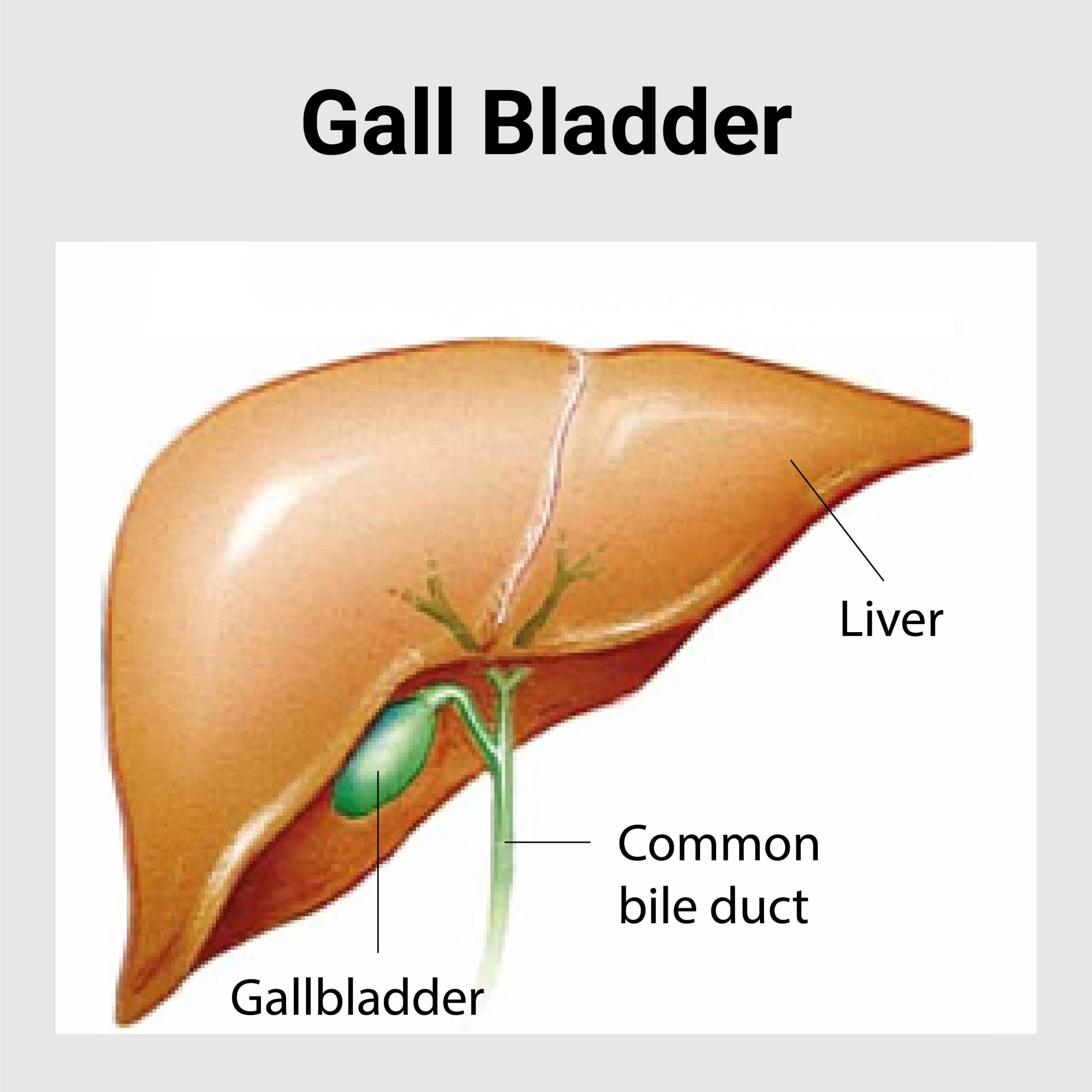
The gallbladder is a small, pear-shaped organ which is adherent to the undersurface of the liver. It stores bile, a fluid that is made in the liver and helps in digesting fat.
When you eat a meal that has fat in it, the gallbladder empties the bile into a tube called the “bile duct.” The bile duct carries the bile into the small intestine to help in the digestion.
- Actue Cholecystitis
- Gall Stone Disease
- Bile Duct Stone
- Choledochal Cyst
- Cancer of Gallbladder
- Cancer of Bile Duct
Acute Cholecystitis
- Cholecystitis means inflammation of the gallbladder.
Causes
- Gallstones
- Tumour
- Bile duct blockage
Symptoms
- Right hypochondriac pain radiating to right shoulder or back
- Abdominal tenderness
- Nausea, vomiting
- Fever
Diagnosis
- USG Abdomen
- CT Scan
Treatment
- Analgesics, Antibiotics
- Surgery- Cholecystectomy
Complications
- Infection
- Gall Bladder perforation
Gallstone Disease
Gallbladder is a small organ located under the liver. Its function is to aid in digestion of food by storing and secreting bile (a digestive juice) into the small intestine when food enters there. Bile is a digestive fluid that is produced by the liver and is made up of several substances including cholesterol, bilirubin and bile salts.
Gallstones are pieces of solid material that form in the gallbladder. Sometimes the cholesterol and pigments present in bile result in formation of hard particles. These stones range in size from tiny sand grains to large ones the size of golf balls.
There are mainly two types of gallstones that can be formed inside gallbladder:
Cholesterol stones: these are the most common type of gallstones, often appears yellow green in color. Almost 80% of the gallstones are cholesterol stones.
Pigment Stones: it is usually formed due to the large amount of bilirubin inside gallbladder. They are dark brown or black in color.
Signs & Symptoms
- Sudden and rapid pain in the upper abdomen and upper back lasting several minutes to few hours
- Back pain between the shoulder blades
- Pain in the right shoulder
- Nausea & vomiting
- Other gastrointestinal problems like – Bloating, indigestion, heartburn, gas etc
Causes
- Gallstone may develop when there is too much cholesterol or bilirubin inside gallbladder secreted by the liver.
- Bile usually dissolves or break down cholesterol however if the liver produces more cholesterols than the bile can dissolve it results in formation of hard stones called cholesterol gall stones.
- Bilirubin is a chemical that is produced when the body breaks down old red blood cells. In some cases such as cirrhosis of the liver and certain blood disorders the liver produces too much bilirubin which contributes to the gallstone formation.
- If the gallbladder often fails to get empty completely the bile may becomes overly concentrated leading to formation of gallstones.
Risk Factors
Following are the various risk factors of developing gallstones:
- Age of above 40 years and female gender: gallstones are more common among women and older people
- Cirrhosis of liver: may cause liver to produce more cholesterol than bile can dissolve which leads to formation of cholesterol gallstones.
- On low calorie diet that leads to rapid weight loss: if a person looses weight too quickly the liver secretes extra cholesterol which may lead to formation of gallstone. Fasting also cause the gallbladder to contract less preventing the gallbladder from emptying completely which may leads to concentrate the gallbladder content and ultimately gallstones.
- Diabetes: diabetic people tend to have higher level of triglycerides which is a type of blood fat that increases the risk of gallstone formation.
- Obesity: this is one of the biggest risk factor. It can cause a rise in cholesterol and also prevent gallbladder from preventing completely.
- Estrogen: it can increase cholesterol and reduce gallbladder motility. Women who are pregnant or who take birth control pills or hormone replacement therapy have higher levels of estrogen and are more likely to develop gallstone.
- Genetics: Having family history of gallstones increases your risk of developing gallstone.
- Cholesterol lowering drugs: they increases the amount of cholesterol in bile increasing the chances of developing gallstone.
Complications
- If the gallstone gets lodged in the neck of the gallbladder it can cause inflammation of the gallbladder called cholecystitis which can cause severe pain and fever.
- Gallstones may get stuck in to any of the tubes that carries bile from gallbladder to small intestine causing blockage of common bile duct. This condition may lead to jaundice or bile duct infection.
- Gallstone may put you at increased risk of developing gallbladder cancer however the chances of gallbladder cancer are usually very small.
Diagnosis
- Abdominal CT Scan: it is a digital imaging test that takes pictures of the liver and abdominal region revealing the presence of gallstone if any.
- Ultrasound: this test uses ultrasonic sound waves to create image of abdominal organs which help find presence of any gallstone.
- Blood tests: to look for amount of bilirubin in the blood and to determine the functioning of liver.
Treatment
- Surgery is often the first choice if there are profound symptoms of gallstone. Laparoscopic gallbladder removal is the most common surgical technique which involves insertion of two to three rod like instrument called laparoscope through tiny holes on abdomen.
- Drugs that dissolve gallstones caused by cholesterol can be used for those who cannot undergo surgery however these drugs may take several years to eliminate the gallstone.
- The risk of developing gallstone can be reduced by adopting healthy lifestyle strategies. Eat balanced diet. Avoid skipping meals. Drink sufficient amount of water every day. Eat fiber rich diet. Maintain healthy weight etc.
Bile Duct Stone
Introduction
Common Bile Duct (CBD) is a tube that carries bile from gallbladder or liver to the small intestine. CBD stone also known as duct stone or choledocolithiasis is a presence of a gallstone in the common bile duct. Gallstone usually forms inside the gallbladder which is a pear shaped organ below the liver. These stone usually remains in gallbladder however sometime it migrates into common bile duct and gets tucked obstructing the passage this condition is called choledocolithiasis or CBD stone.
Signs & Symptoms
CBD stones may not have any signs & symptoms for months or even years. However, if the blockage becomes severe, the following signs & symptoms may be experienced:
- Pain in the upper or middle part of abdomen
- Fever
- Jaundice
- Loss of appetite
- Nausea & vomiting
Causes
- Gallstone may develop when there is too much cholesterol or bilirubin inside gallbladder secreted by the liver.
- Too much cholesterol: Bile usually dissolves or break down cholesterol however if the liver produces more cholesterols than the bile can dissolve it results in formation of hard stones called cholesterol gall stones.
- Too much Bilirubin: Bilirubin is a chemical that is produced when the body breaks down old red blood cells. In some cases such as cirrhosis of the liver and certain blood disorders the liver produces too much bilirubin which contributes to the gallstone formation.
- Poor emptying of Gallbladder: If the gallbladder often fails to get empty completely the bile may becomes overly concentrated leading to formation of gallstones.
Risk Factors
Following are the risk factors of developing CBD stone:
- Obesity
- Prolonged fasting
- Rapid weight loss
- Lethargic lifestyle
- Pregnancy
- Age & Gender: older people and women are more likely to have CBD stone
- Family history of gallbladder disease
Complications
- Acute Cholangitis: CBD stone can cause infection in the bile. The bacteria from the infection can spread rapidly. It can move into the liver or Ductal system. It can become a life threatening infection
- Hepatic Abscess
- Billiary cirrhosis
- Pancreatitis
Diagnosis
- Abdominal CT Scan: it is a digital imaging test that takes pictures of the liver and abdominal region revealing the presence of gallstone if any.
- Ultrasound: this test uses ultrasonic sound waves to create image of abdominal organs which help find presence of any gallstone.
- Endoscopic Ultrasound (EUS): an ultrasound probe is inserted on a flexible endoscopic tube and inserted through mouth to examine the digestive tract.
- Endoscopic Retrograde Cholangiography (ERCP): it is a procedure used to identify stones, tumors and narrowing in the bile ducts. An endoscopic tube is inserted through the mouth and dye is injected into the ducts where they can be visualized with x rays.
- Magnetic Resonance Imaging (MRI): MRI scans of the gallbladder, bile ducts and pancreatic duct called Magnetic Resonance Cholangiopancreatography (MRCP).
- Blood tests: to look for amount of bilirubin in the blood and to determine the functioning of liver.
Treatment
The treatment of CBD stone includes one or more of following:
- Stone extraction
- Lithotripsy: breaking the stone into small pieces allowing them to pass through
- Cholecystectomy: surgery to remove gallbladder and stone
- Sphincterotomy: surgery to make a cut into the common bile duct to remove stones or help them pass
- Biliary stenting
- Laparoscopic CBD exploration
- Endoscopic Sphincterotomy is the most common treatment of CBD stone. During this procedure a balloon or basket type device is inserted into the bile duct and used to extract the stones.
Lifestyle changes such as physical activity and dietary changes such as increasing fiber and decreasing saturated fats can reduces the chances of developing CBD stones.
Choledochal Cyst
a congenital dilatation of part or whole of the bile duct.
Symptoms
- Intermittent abdominal pain
- Right upper abdominal mass
- Jaundice
Diagnosis
- USG Abdomen
- CT Scan
- MRCP,ERCP
Treatment
- Surgery and removal of the disease part
Cancer of Gallbladder
American Indians and Hispanics. If it is diagnosed early enough, it can be cured by removing the gallbladder, part of the liver and associated lymph nodes. Most often it is found after symptoms such as abdominal pain, jaundice and vomiting occur, and it has spread to other organs such as the liver. The incidence of gall bladder cancer is increasing in China as well as north central India.
It is a rare cancer that is thought to be related to gallstones building up, which also can lead to calcification of the gallbladder, a condition known as porcelain gallbladder. Porcelain gallbladder is also rare. Some studies indicate that people with porcelain gallbladder have a high risk of developing gallbladder cancer, but other studies question this. The outlook is poor for recovery if the cancer is found after symptoms have started to occur, with a 5-year survival rate close to 3%.
- Gender: twice more common in women than men, usually in seventh and eighth decades.
- Obesity increases the risk for gallbladder cancer.
- Chronic cholecystitis and cholelithiasis.
- Chronic typhoid infection of gallbladder. Chronic salmonella typhi carriers have 3 to 200 times higher risk of gallbladder cancer than non-carriers and 16% lifetime risk of development of cancer.
- Various single nucleotide polymorphisms (SNPs) have been shown to be associated with gallbladder cancer. However, existing genetic studies in GBC susceptibility have so far been insufficient to confirm any association.
Signs and symptoms
- Steady pain in the upper right abdomen
- Weakness
- Loss of appetite
- Weight loss
- Jaundice and vomiting due to obstruction
Early symptoms mimic gallbladder inflammation due to gallstones. Later, the symptoms may be that of biliary and stomach obstruction.
Disease course
Most tumors are adenocarcinomas, with a small percent being squamous cell carcinomas. The cancer commonly spreads to the liver, bile duct, stomach, and duodenum.
Diagnosis
Early diagnosis is not generally possible. People at high risk, such as women or Native Americans with gallstones, are evaluated closely. Trans-abdominal ultrasound, CT scan, endoscopic ultrasound, MRI, and MR cholangio-pancreatography
(MRCP) can be used for diagnosis. A biopsy is the only certain way to tell whether the tumors growth is malignant or not.
Treatment
The most common and most effective treatment is surgical removal of the gallbladder (cholecystectomy) with part of liver and lymph node dissection. However, with gallbladder cancer’s extremely poor prognosis, most patients will die by one year following the surgery. If surgery is not possible, endoscopic stenting of the biliary tree can reduce jaundice and a stent in stomach may relieve vomiting. Chemotherapy and radiation may also be used with surgery. If gall bladder cancer is
diagnosed after cholecystectomy for stone disease, reoperation to remove part of liver and lymph nodes is required in most cases. When it is done as early as possible, patients have the best chance of long term survival and even cure.
Cancer of Bile Duct
Causes
- Certain diseases of the liver or bile ducts- Primary sclerosing cholangitis ,Bile duct stones, Choledochal cysts,Liver fluke infectionsCirrhosis,Infection withhepatitis B virus or hepatitis C virus
- Inflammatory bowel disease
- Older age
- Obesity
- Family history
- Diabetes
- Alcohol
- Smoking
- Pancreatitis (inflammation of the pancreas)
- Infection with HIV (the virus that causes AIDS)
Symptoms
- Abnormal liver function tests,Jaundice
- Abdominal pain
- Generalized itching
- Weight loss
- Fever
- Changes in stool or urine colour
Diagnosis
- Blood Test
- Biopsy
- CT Scan
- ERCP
Treatment
- Surgery to remove the bile duct, and sometimes part of the liver
- Chemotherapy
- Radiation
- Stent placement
Complication
- Liver Failure
- Pneumonia
HOSPITAL ADDRESS
132ft. Ring Road, Helmet Circle, Memnagar, Ahmedabad – 380052. Gujarat, India.
EMERGENCY( 24X7 )
Mobile: +91 – 99047 44410
Help Line : +91 – 98244 40044
+91 – 79 – 2791 4444
E-MAIL ADDRESS
contact@xdemo.app
WHY KAIZEN ?
With a vision to extend World Class healthcare solutions to the community through advances in medical technology, medical research and by adopting best man power management practices , Kaizen hospital was established in Ahmedabad in 2011.

